Reflecting on guidelines and departmental disparity in protocols for suspected physical abuse in children


In 2008, the Royal College of Radiologists (RCR) published standards for radiological investigations of suspected non-accidental injury which was updated in 2017 with several modifications (1). Published in conjunction with the Royal College of Paediatrics and Child Health and the College of Radiographers, the aim of the standards was to provide a consistent nationwide approach to imaging with guidance for both radiologists and radiographers to ensure that correct, timely and appropriate imaging is obtained which would stand up to legal scrutiny.
Whilst the standards provide clear instructions for referral pathways and the images which should be taken, they are less specific about matters such as the level of further training radiographers and radiologists should have regarding experience and safeguarding issues. The document contains information about the range of imaging modalities that may be employed, but not necessarily the importance of them and when they should be utilised. Each case should be assessed on an individual basis with positive or suspected findings having follow up or further imaging which are considered appropriate for that particular patient and presentation. For larger centres with clinicians who have expertise of paediatric injuries and their radiological presentation this is not a problem, but smaller general imaging departments which encounter such patients may not always appreciate very subtle abnormalities, what further imaging may be warranted and the significance of these anomalies.
Early identification

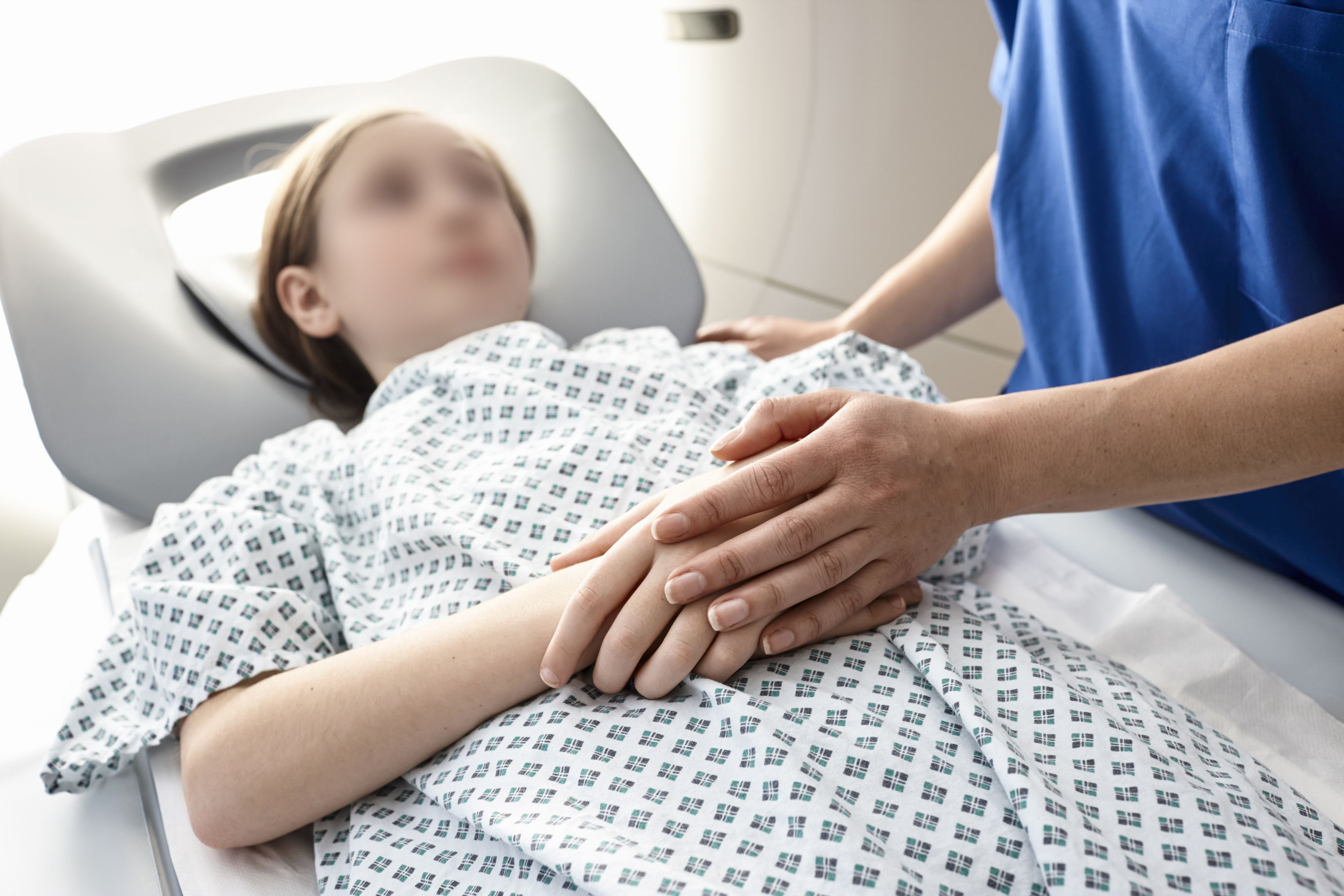
It is important that a child receives the correct diagnosis, whether investigations prove positive or negative. Imaging is often central and important evidence when considering the removal of a child from their family to be placed into care, or any subsequent court proceedings taken against the guardians. There have been many high-profile cases which have highlighted the importance of identifying vulnerable children on their first and every interaction with health and social care. It is therefore imperative that any pathology present at the time of the skeletal survey is demonstrated and appreciated correctly as trauma or other metabolic or skeletal conditions. Similarly, it is necessary that a negative examination be recognised as such and that normal variants are accurately identified.
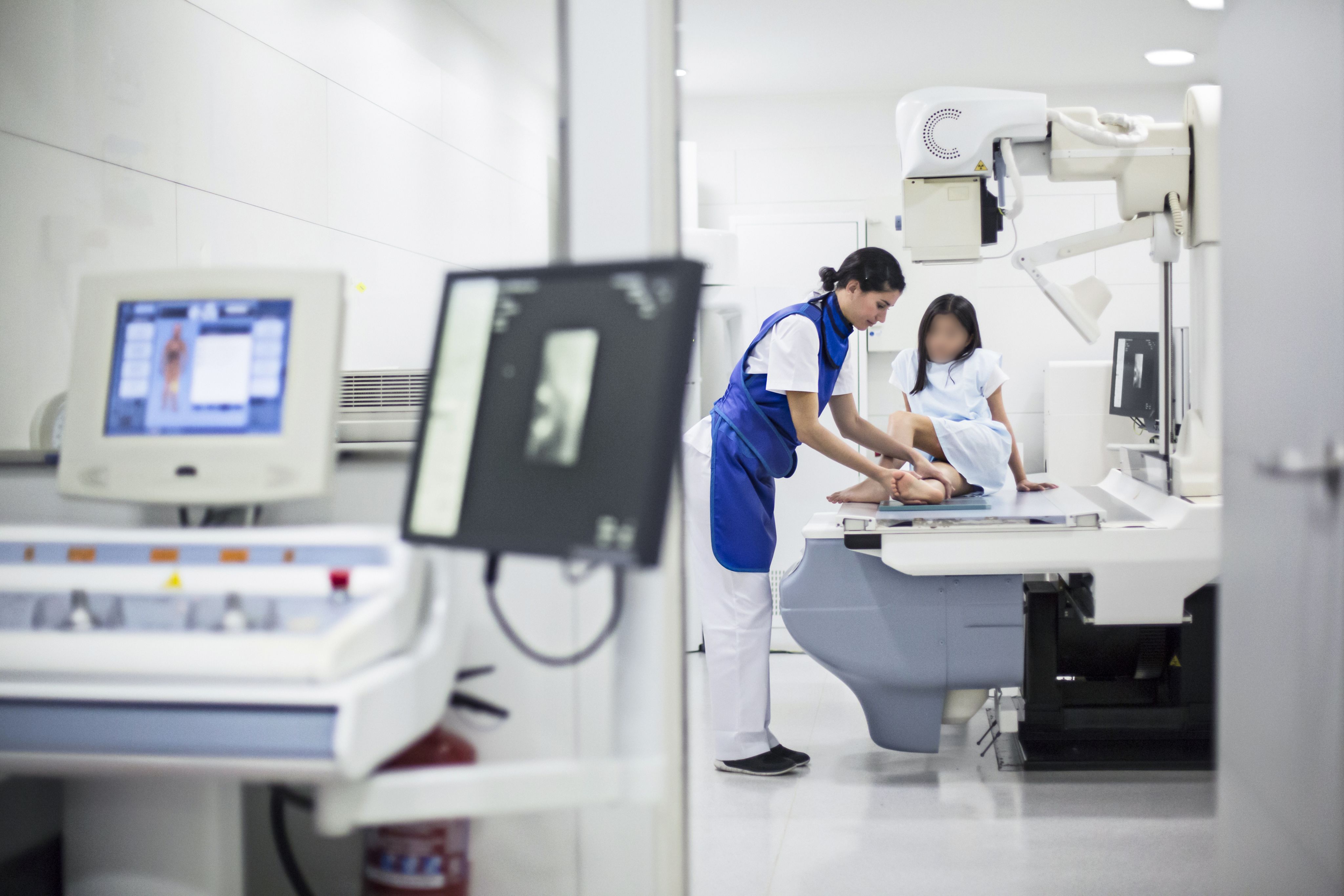
As the full initial skeletal survey involves at least 23 exposures and by necessity involves irradiation of particularly radiosensitive organs there is always a concern that the dose should be kept to a minimum whenever possible. However, this has to be offset by the need to obtain high quality images with optimum sensitivity and specificity. The ‘as low as reasonably practicable’ (ALARP) principle should always be applied with radiographers using all the tools of best practice to keep the radiation dose for each image as low as possible. Modern, well-maintained X-ray equipment is capable of producing high resolution images with lower doses than older machines. Accurate collimation is important as this helps with dose reduction and image quality, however the importance of including the whole area of interest as well as anatomical side markers can limit this at times.
Studies have shown that despite more images being included in the survey than previous guidelines have indicated, the overall radiation dose to the patient remains within acceptable limits with typical doses equivalent to approximately one month of background radiation (2).
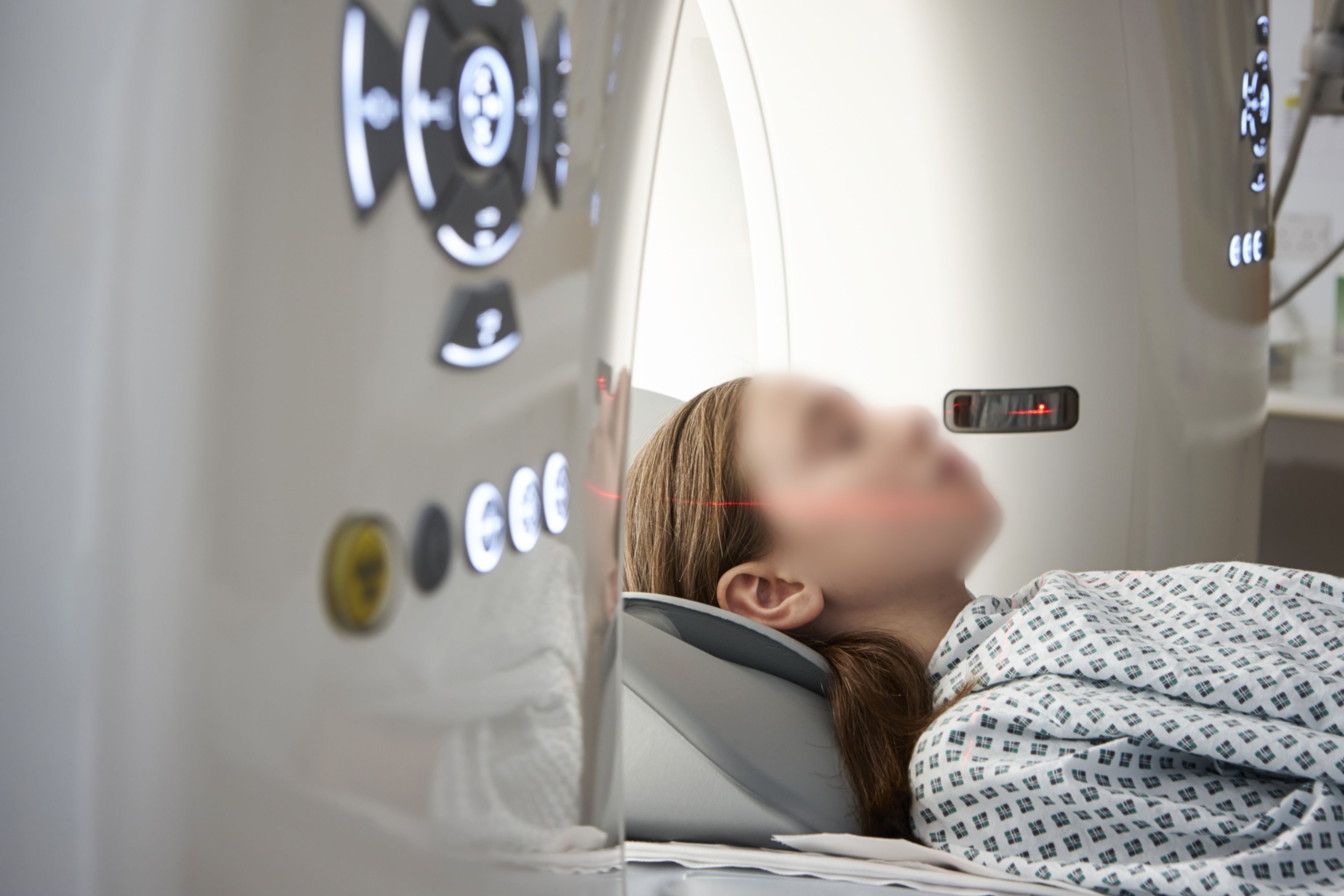
It is recognised that there is sometimes a reluctance to include Computed Tomography (CT) head scanning as part of the investigation, most probably when there is a low suspicion of any trauma or there are no neurological symptoms (3). However, any injury to the skull may result in an associated brain injury with more serious consequences and must be ruled out. The latest scanners deliver faster scan acquisition times, which reduces the likelihood of movement artefact with lower patient dose Therefore, the risk versus benefit consideration in conjunction with the guidelines favours the inclusion of a CT head and eliminates the need for two skull images from the skeletal survey.
A number of retrospective audits have been carried out which have investigated the value of the follow up survey (4). Although this includes fewer images than the initial survey, it has been found to be beneficial as it can detect injuries which were not shown on the initial survey (5).

It might be considered that projection radiography is not always the best imaging modality to detect very subtle or occult fractures, particularly metaphyseal injuries and that utilisation of Magnetic Resonance Imaging (MR) and ultrasound may be useful. However, the cost of such imaging and the difficulties associated with accessing these services will determine whether they are employed on a case-by-case basis according to the level of clinical suspicion. These cannot therefore be included in any definite way in the RCR guidelines other than to acknowledge their availability (6).
Guidance on projection imaging is comprehensive and clear as to which images should be taken, how soon after the referral is made and when the double report should be issued. Although the best scenario would be to complete the examination and issue a final report within 24 hours, there is flexibility within the guidelines to postpone the examination until the necessary staff are available to undertake the procedure. This is particularly true when a request for imaging is made outside of normal working hours.
Delays and misdiagnosis
Potential problems and delays may arise when the child is too unwell to attend, or other clinical concerns take priority. This should be documented in the patient’s notes and radiology files.
A delay in obtaining a double read report might lead to a delayed or misdiagnosis. It has been found that there could be a discrepancy in interpretation of findings of up to 4.5% (7). The guidelines recommend that both reports are written by radiologists with at least six months of specialist paediatric training with experience of suspected physical abuse and suggest that the reports should be in agreement. Any difference of opinion should therefore be discussed, and a third expert opinion sought if appropriate even though this will ultimately lengthen the interval between imaging and release of the final report.
With any team providing a specific service, there is always the balance between how many people to include, ensuring there is likely to be sufficient appropriate personnel available when needed against having so many so that experience and involvement is limited. Many radiology departments within local secondary care centres are currently under significant work pressures requiring radiographers to work long hours including weekends and unsocial hours. This results in more limited availability of appropriately trained radiographers during the limited time frames stipulated in the guidance.
The training required for radiographers involved is vague. The RCR guidelines say that it should be ‘documented education and training’ with ‘level 3 knowledge skills and competence’ referring to the ‘Safeguarding children and young people: roles and competences for health care staff’ (8). This document however contains information about agencies set up to protect children, safeguarding concerns to be aware of and a knowledge of forensic and legal matters but nothing specific for radiographers and radiologists who encounter these children for a short but intense period of time with a specific role.
The physical acquisition of high-quality images depends on the ability of the individual radiographers. As examinations are most often carried out on live subjects, the guidance from the Society of Radiographers on forensic radiography is clear that radiographers have a duty to adhere to all the usual codes of conduct and relevant regulations for diagnostic imaging, engaging safe practice and efficient use of non-ionising radiation (9).
Additional guidance

Imaging for suspected physical abuse comes under a broader umbrella of forensic radiology but there is no reference to this in the RCR guidelines. More detailed information concerning the forensic aspects is provided in the Guidance for Radiographers providing Forensic Radiography Services document, published by the Society of Radiographers in 2014, which is helpful to radiographers, radiologists and departmental managers alike (9).
The guidance for forensic radiography also stipulates that radiographers must be educated and trained at postgraduate level and receive regular updating as part of their continuing professional development (CPD). It provides more detailed information for radiographers and radiology departments regarding storage of examinations and ensuring that appropriate witnesses and documentation is appropriate and protected so that it will be admissible if required by safeguarding agencies or a court of law. Radiology departments should comply with the provisions as set out in the relevant codes of professional conduct and with all relevant regulations for diagnostic imaging and the safe and efficient use of ionising and non-ionising radiation.
Radiographers carrying out forensic examinations must comply with the local protocols for forensic imaging. These protocols are required to contain specific information about referral rights and pathways, confidentiality for the patient and their families and in particular, how continuity of evidence and the presence of an appropriate witness is maintained.
It is also recommended that radiographers should write their own record of the event for future reference if required, although few radiographers have had help with how this might be done other than experience of writing reflective accounts of work occurrences as part of their CPD. This might for example, include noting unusual behaviour or interaction between patient and carer, accusations made against others not present and could take any form considered appropriate by the imaging department involved but should be kept securely with all the other documentation. Any further visible signs of trauma not previously recorded, which become apparent at the time of imaging must also be documented and the information passed on to the clinical team immediately after the imaging is completed.
The impact of undertaking these examinations is difficult to appreciate as different clinical centres will encounter these scenarios more or less frequently than others and have their own locally arranged setup in place which relies upon many or few radiographers and radiologists to complete them, not only accurately but within a short period of time. This can take its toll on those involved, particularly when the case is complex or high profile, and it is appreciated at the time of imaging that the examination will play a vital role in determining the future welfare of the child (10).
The guidance for radiographers providing forensic radiography services includes a section about the welfare of radiographers. Employers have a responsibility to minimise the risk of psychological harm to employees but many imaging departments do not have the resources to support staff following any difficult case, not necessarily those of suspected physical abuse but hopefully this will improve in the future. One of the positive aspects to come about following the Covid-19 pandemic is that healthcare settings and many other employers are more aware of the importance of good mental health for everyone and are more familiar with where and how to access help.
Ensuring radiology compliance with RCR guidelines for suspected physical abuse is imperative for maintaining the reliability of radiographic evidence depended on by safeguarding and legal teams. Ongoing training, addressing knowledge gaps, and fostering effective team dynamics are essential components of achieving this compliance. Moreover, the number of radiographers and radiologists within the team plays a pivotal role in the successful implementation of these guidelines and wellbeing support of all involved as required.
There are some recently published guidelines from the Society of Radiographers which include forensic examination of the living which are worth reviewing.
References
1.The Royal College of Radiologists and Society and College of Radiographers. The Radiological Investigation of Suspected Physical Abuse in Children, revised first edition. London: The Royal College of Radiologists; 2018.
2. Rao R, Browne D, Lunt B et al. Radiation doses in Diagnostic Imaging for Suspected Physical Abuse, Archives of Disease in Childhood, 2019, 104:863-868.
3. Halstead S, Scott, G. Thust, S, Hann G. Review of the new RCR guidelines (2017): The radiological investigation of suspected physical abuse in children, Arch Dis Child Educ Pract Ed 2019; 104 309-312.
4. Bennett BL, Chua MS, Care M, Kachelmeyer A, Mahabee-Gittens M. Retrospective review to determine the utility of follow-up skeletal surveys in child abuse evaluations when the initial skeletal survey is normal. BMC Res Notes. 2011 Sep 12;4:354.
5. Kleinman PK, Nimkin K, Spevak MR, Rayder SM, Madansky DL, Shelton YA, Patterson MM. Follow-up skeletal surveys in suspected child abuse. AJR Am J Roentgenol. 1996 Oct;167(4):893-6.
6. Lawson M, Tully J, Ditchfield M, Metcalfe P, Qi Y, Kuganesan A, Badawy M. A review of current imaging techniques used for the detection of occult bony fractures in young children suspected of sustaining non-accidental injury. Journal of Medical Imaging and Radiation Oncology, 2022, 66, 68-78.
7. Nguyen A, B Rad Med Imag (Hons) Hart, R PhD Imaging of non-accidental injury; what is clinical best practice? First published: 24 March 2018.
8. Royal College of Paediatrics and Child Health. Safeguarding Children and Young People: Roles and Competencies for Healthcare Staff. Fourth edition: January 2019 www.Safeguarding children and young people - roles and competencies | RCPCH Accessed October 2023.
9. Society and College of Radiographers. Guidance for Radiographers providing Forensic Radiography Services. 2014. Guidance for Radiographers Providing Forensic Radiography Services | SoR www.sor.org/learning-advice/professional-body-guidance-and-and-publications/documents-and-publications/policy-and-document-library/guidance-for-radiographers-providing-forensic-radiography Accessed October 2023.
10. Eaton J. A suspected physical abuse examination: the radiographer’s perspective, Journal of medical Imaging and Radiation Sciences, 2023, 54 S6-S7.