


Spotlight on CPD:
The latest in clinical imaging e-learning
Dorothy Keane, clinical lead for the e-learning for healthcare Clinical Imaging programme, updates radiographers on the latest developments

Dorothy Keane
Dorothy Keane
There are over 112,000 enrolled learners on the Clinical Imaging Programme, and 50,800 active regular users. Around half are radiographers and student radiographers, the rest are doctors, other allied health professionals and nurses.
There are almost 500 sessions in the Clinical Imaging programme. These sessions are split into chapters, which include knowledge checks, interactive learning opportunities, guidance and resources.
Dorothy said: “You'll see almost every topic, every area of imaging covered in our programme. If you haven’t accessed them, do. They’re really useful sessions.”
New content
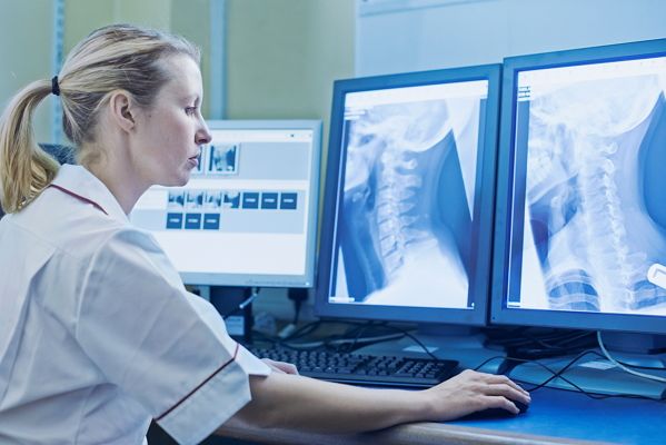
The SoR recently launched the Adult Pathology module to enable recognition of bone or soft tissue changes on conventional radiographs. These sessions help you recognise a range of pathologies from arthritis to gout, to malignant tumours. Our current development includes Paediatric Pathology, the importance of which is evident; identifying a malignant tumour early could save a life.
The Society has also published material which will support radiographers recognising pulmonary emboli in CT. Other new content includes supporting overseas radiographers, an introduction to AI and inclusive practice in medical radiation safety.
Last year saw the addition of learning modules for IV cannulation, osteoporosis and fragility fractures and the team has also recently worked with radiographers to develop sessions to help with understanding accessory projections.
Orthopaedic sessions were added because of a high volume of requests from reporting radiographers, saying that so many new implants had been developed in orthopaedics they were unsure how to describe them and their appearances when complications occur.
Dorothy added: “In the last two years, the programme has diversified slightly from image interpretation and from purely clinical work. Sessions on pathology and the identification and description of radiography are still in development, but other groups are now working to develop guidance for such topics as inclusive practice in medical radiation safety and an introduction to artificial intelligence.”

Background
The initiative began with sessions on adult and paediatric skeletons and how to interpret them, helping radiographers understand the anatomy, mechanisms of injury, before moving on to fractures and basic pathology.
Further modules include cross-sectional imaging in neuro-emergencies and topics focused on technology. These modules cover the different types of imaging, how they work and how radiographers can improve the way they are used.
The ultrasound curriculum in its entirety, including obstetric and general, is in the process of being updated for a second time.
Clinical Imaging also includes sessions on forensic radiography and signs of suspected physical abuse.
Other modules include sessions on CT Anatomy, MRI, GI-GU, cardiac and neurological imaging. The sessions are developed by the NHSE e-learning team and radiographers across the country, who have developed specialist knowledge through their own further education and experience.
Sessions can also be used as continuing professional development. Radiographer feedback shows that topics such as dementia, working with children, consent and dignity have been combined with departmental talks from clinical staff from other departments to reinforce subject matter and support CPD.
Dorothy Keane
Dorothy Keane
Find out more...
Dorothy Keane MBE joined the Society of Radiographers as the clinical lead for the Image Interpretation (now Clinical imaging) programme in 2010. Prior to that she worked as a consultant radiographer and developed a system in which radiographers could provide provisional clinical evaluations on all conventional radiographs.
For 10 years, Dorothy worked to develop and audit that process, learning how to educate radiographers who have provided feedback about what type of training they required, this feedback and development served as the foundation for the entire Clinical Imaging programme.
In 2018, Dorothy was name in the New Year Honours List, and was awarded an MBE for her services to the profession.
Find out more about the Clinical Imaging programme here.
Image credit: Pakin Songmor/Getty Images