Professional
15 Minutes With...
Steven Edwards
Veterinary MRI radiographer


Radiography roles take many different shapes, and not all of them involving humans. Steven Edwards talks Synergy through a day in a veterinary hospital
Can you give us a little background on your experience?
It was a huge change going from a busy NHS hospital, imaging the general public, to imaging animals at a specialist referral veterinary hospital. I thought I’d be bitten far more working with animals than people, how wrong I was.
I have been qualified since 2008 and commenced my career working at New Cross Hospital in Wolverhampton. I started in X-ray and loved the experience. I threw myself into trauma as much as possible, enjoying adaptive techniques and found A&E work suited me. After 18 months I got a post in the cath labs where I stayed for another four years. I left in 2013 to learn MRI with Alliance Medical.
Mobile life presented a lot of different pressures, which I enjoyed. I learnt the basics, but wanted more complex scanning, so headed back to New Cross Hospital. I started in MRI and quickly learnt cardiac MRI, which was my favourite aspect of the job. I really enjoyed the different challenges it presented. The cardiologists I worked with always talked through the pathology and I find that makes scanning easier and more interesting. I then took an advanced practice role, which I did for a number of years until I found a post working with animals at the Dovecote Veterinary Hospital in Castle Donington, Leicestershire.
What made you want to become a veterinary radiographer?
I wasn’t aware of specialist veterinary hospitals and happened to come across a job advert for this post online by chance. It was daunting to go from an advanced practitioner in MRI, scanning people, a job I knew inside out, to working in a field where very few radiographers have the chance to encounter. There are plenty of challenges, but the rewards are fantastic. Who wouldn’t want to be playing with puppies at 9am on a Monday morning?
It isn’t all fun and games though. It is very demanding, as I normally see the complex cases that cannot be seen by a local vet. I mainly image neurology cases. For example, if a dog has a disc extrusion, brachial plexus tumours or a brain tumour, I perform a range of sequences including specialist scans like spectroscopy or diffusion tensor imaging.
What does a typical day look like as a veterinary radiographer?
The day starts at 8am where I prep the scan room. This means I fill up the isoflurane tanks with anaesthetic and ensure the circuits are connected and working. I make sure to have the correct sizes of cuffs for monitoring, and stock for the list.
From here I go to X-ray and help take the pre-op films for orthopaedic cases, before the MRI list starts. Once the first case for MRI is in the practice, I will help to hold the animal for a cannula and put together fluids for the case. The vet will complete a safety questionnaire with the owners and discuss with me. The nurse and vet will anaesthetise the animal and we go straight into MRI. Once the list is over I send the images for reporting and tidy up. We have emergencies 24/7 so we have to be ready at a moment’s notice. Normally lists finish around 4pm, allowing for the clean-up. Hair is very difficult to get out of the scanner in the volumes we see.
My role isn’t just standard MRI radiographer. Much like roles in the cath labs you are part HCA, part nurse and a radiographer. This is why I enjoy the role so much: variety and pressure. I have been doing this for 18 months and I can say I have a greater understanding of the patient pathway now more than I ever had in the NHS.
This is solely due to seeing the whole patient journey in a day, from consultation to imaging and treatment. In my day I can be seen in all departments. I will regularly clean or help hold to cannulate. I started to take X-rays again, and after nearly a decade out of plain film and using a stationary tube, I can say it is a challenge.
Most radiographers would be shocked at the practice of how we position and the aids we have. The imaging table has loops and cleats to tie limbs to for imaging. It isn’t as if you can just ask the cat to stay in this position. My first X-ray for a decade was on a rabbit for a mandible fracture and I would say that was advanced adaptive techniques. Just like in the human world, getting a lateral knee/stifle is an art form. It is essential to destress outside of work as we do see the worst cases scenario. The same dark humour that is in the human hospital is in the veterinary world as a coping mechanism. It’s a great way to help with the pressures of what you deal with on a daily basis.
Every day is different so I’ll share a random day which started with an emergency. A Dachshund had become paraplegic, with pain response, and required to be seen by the specialist neurologist. They were rushed into the hospital and taken straight to MRI finding a disc extrusion at L4/L5. The case was then taken to theatre for a hemi-laminectomy to remove the disc material causing the compression on the cord and nerves.
The pre-booked list then started with a domestic shorthair cat with vestibular signs. They arrived with head tilt and suspected otitis media, inflammation or infection of the middle ear. This is treatable with antibiotics and steroids. We proceeded with MRI and using T2/Flair/T1 sequences I found there was huge inflammation in the inner ear, with the tympanic bulla being full of fluid. Then a Border Collie came in with back right lameness and ataxia, sudden onset after exercise. We took this into the scanner and found it had had a fibrocartilaginous embolism, which is a spinal stroke. It was a late lunch type of day as another emergency came through. A French Bulldog off his hind legs, and after finding a huge disc extrusion at T12/T13, was taken to theatre.
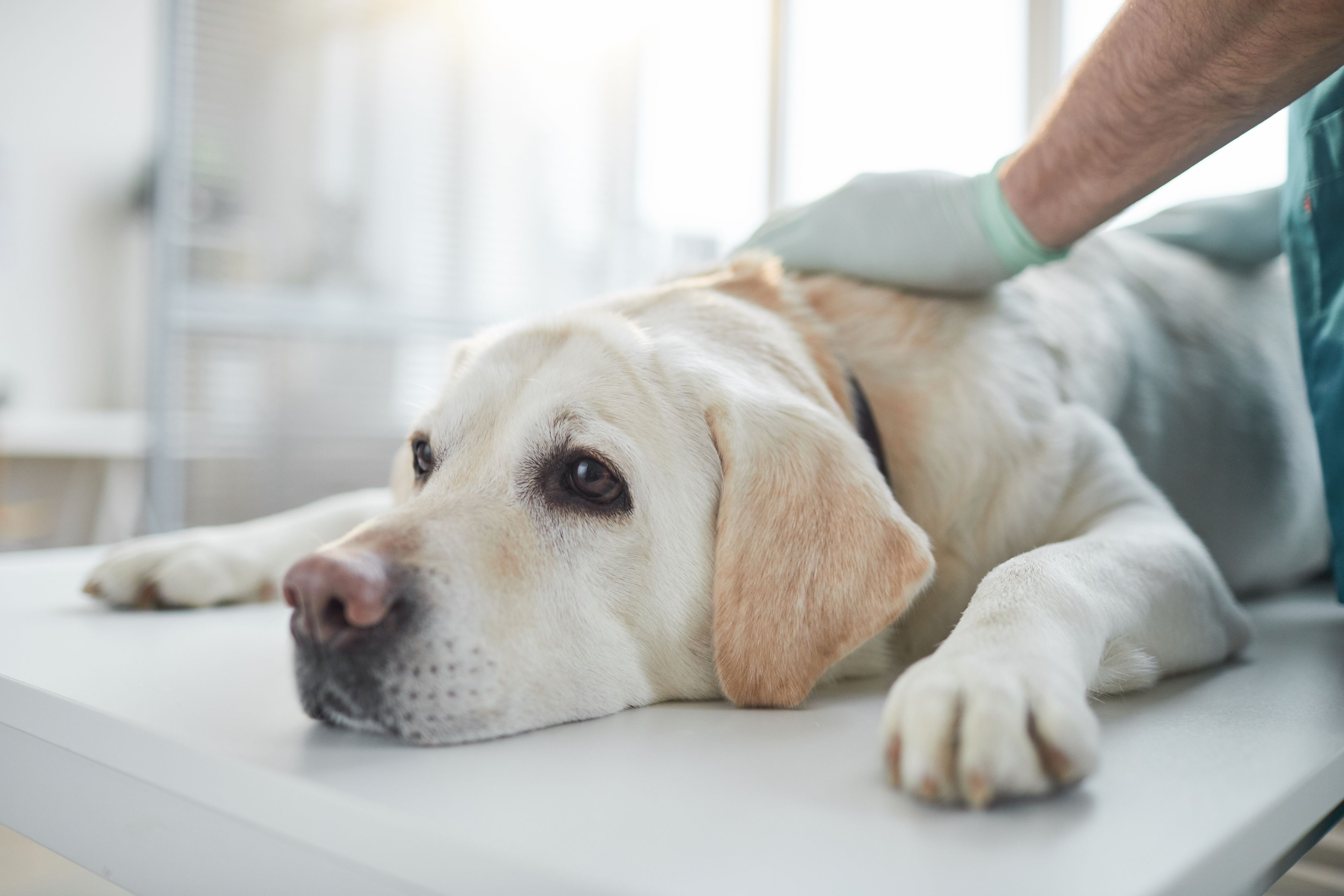
The final case of the day was epilepsy in a Cavalier King Charles Spaniel, which I scanned as a routine brain protocol. There was no pathology seen and was diagnosed with idiopathic epilepsy, requiring only medical treatment. Not all days are so busy. Some days I will take a Labrador into X-ray to image its stifle before the surgeon performs a tibial plateau levelling osteotomy or a chest X-ray for a case with breathing issues.
What, for you, are the best parts of being a veterinary radiographer?
Each case will involve the radiographer, a nurse, and a vet. The vet usually stays for the entire case. There are lots of discussions on each case, and my opinion is very much valued. Each case is very unique in the way that we have routine sequences but regularly we add sequences to highlight pathology such as 3D nerve root imaging. I would say 90 per cent of cases will have significant pathology, which can require immediate treatment. This is a huge contrast to the NHS work.
The scanning side of things can be very challenging. The scanner I use is for a human so every protocol has to be adapted. A large German Shepherd brain is relatively straightforward as the anatomy is similar in size to humans, but a 4kg Chihuahua can be a challenge. This is due to the lack of signal. When scanning with protocols from the scanner they have to be cut down hugely to cater for the tiny field of view (FOV) required. A Chihuahua brain can be as small as 3.5 cm, so using a 5mm slice would not produce a diagnostic image. We push the scanners to the limit of resolution versus FOV.
This can increase scan times drastically so I have to be good at cutting back where I can to balance time versus diagnostic image quality. It is not uncommon to have a seven minute sequence in the brain. Likewise when scanning a German Shepherd thoraco-lumbar spine I have to boost the signal by increasing FOV and averages due to the size of the spinous processes. The signal drop off is an issue here due to the depth of the spinal cord. You have to be comfortable manipulating protocols and making sequences from scratch. Every animal is slightly different, so you cannot just have a set protocol.
We use very thin slices for our neuro work, and even a large breed’s brain is dramatically smaller than that of a human. I see a lot of advanced pathology because animals adapt very well until it gets to a severe stage where you notice symptoms. We do have lists each day but we also take emergencies: sometimes one case, sometimes four or five. Each additional case will have a 30-minute consult, a one-hour MRI, and then up to three hours in surgery. It's great to see the full pathway of each case. For example a French Bulldog coming in paraplegic, having neurological tests, scans, surgery and post-op care, and within 48 hours they are going home.
What advice would you give to those just starting their radiography journey?
If I was to give advice to students or newly qualified staff it would be to look out for interesting roles. You don’t have to stick to the same career pathway as everyone else. Go after what you find interesting. Radiography has a huge scope, not just within the NHS. I do not regret my choices and have enjoyed all of my posts. I have only just started in this role and I’m constantly learning and adapting. Maybe in the next five years we will see more of these specialist animal facilities popping up, requiring more and more advanced imaging. This is great for the profession and creates a new branch for people to learn. Love what you do and enjoy the team you work with.


Steven with a patient
Steven with a patient

The X-ray table
The X-ray table


Paraplegic Dachshund (1)
Scan of the paraplegic Dachshund


Paraplegic Dachshund (2)
Scan of the paraplegic Dachshund

Chihuahua
Scan of a Chihuahua

Steven looking over scans
Steven looking over scans
Where I work...
A closer look at the workspace of a veterinary radiographer
Consulting rooms
This door to the consult rooms leads directly into the prep area, with theatre/CT/ X-ray/US and MRI leading off it
The drug store
Our store space for drugs, syringes, and clinical stock
The tub sink
Tub sinks used for washing dogs and scope procedures
Piped gases
These are our piped gases and isoflurane vaporiser, used for anaesthesia
Anaesthetic station
One of our three anaesthetic stations with its own isoflurane vaporiser, a table for the animal, and a cannulation trolley
Find out more...
Steven Edwards is a diagnostic radiographer based at the Dovecote Veterinary Hospital in Leicestershire.
If you are a radiographer with an interesting role and would like feature in a '15 minutes with...' article contact alexander.ballinger@wonderly.agency.
Image credits: Dovecote Veterinary Hospital
SeventyFour/ iStock / Getty Images Plus